Showing posts sorted by relevance for query tb. Sort by date Show all posts

Tuesday, September 30, 2008

Day #92 - "I don't know"

Wow. What a great case. It isn't often that I truly throw my hands up and say that "I don't know" but this was one of them.

But I hope the exercise in reasoning was useful for you. I think we demonstrated an approach that was safe, cost-effective, and broad in going through the history and physical exam.

Dr. Gold previously spoke about meningitis (bacterial) and that talk is available here.

In approaching a patient with multiple infarcts we need to consider the following differential:
  • Emboli - Arising from the heart or great vessels. One possibility is subacute bacterial endocarditis, particularly given the history of night sweats. I think this patient needs blood cultures drawn with blind subculturing (endocarditis blood cultures) to look for unusual pathogens like brucella. I also think he needs a trans-esophogeal echo looking for a cardiac source including a bubble study to exclude PFO. This can also look at the aortic arch.
  • Ischemia -- Either bad luck from uncontrolled risk factors, or genetic stroke syndrome like CADASIL, Fabry's or MELAS, or thrombophilia like antiphospholipid antibody syndrome, ATIII deficiency or hyperhomocysteinemia. I would exclude thrombophilia. As a last resort I would look into genetic testing. Night sweats don't fit with ischemia.
  • Vasculitis (Night Sweats would fit with this too)
  • Other
If the echo is clean, I would consequently perform angiography (MRA possibly conventional) in this patient and look for evidence of vasculitis. I think given the night sweats and epidemiology that TB needs to be excluded prior to high dose steroids. I would repeat the lumbar puncture to ensure that it is still acellular (early TB can be acellular) and again send TB studies.

I would confirm whether or not he has had a mantoux. He should have one prior to immunosuppression anyways and should be treated for latent TB if this does not represent CNS TB.

I would probably scan his chest/abdomen/pelvis to look for evidence of malignancy, lymphoma or TB disease -- or something safer than the brain to biopsy and make a diagnosis.

Ultimately he may require a stereotactic brain biopsy to exclude TB and make a diagnosis. In a young man, with progressive infarcts and no other cause, I would discuss this with the patient.

TB is treatable, thrombophilia and emboli preventable, and lymphoma may be cured but CADASIL can not so we had better be sure!

Wednesday, January 6, 2010

TB - Tuberculosis Hodge-podge

Yesterday we saw an interesting case at the end of the day of a patient on immunosuppression who presented with a new left sided pleural effusion and some parenchymal changes compatible with a flare of her underlying lung disease vs. tuberculosis.

This patient comes from an endemic country and has evidence of fibronodular changes on chest x-ray (RR for reactivation ~15). There has been no previous TB treatment.

This highlights a large number of good teaching points:

  1. Diagnosis and management of latent TB.
  2. Diagnostic approach to possible TB pleuritis.
  3. The interplay between TB and TNF-alpha inhibitors.
  4. The radiographic manifestations of TB and the differences in immunosuppressed hosts.
  5. The yield of bronchoscopy in patients who are not coughing.
  6. The potential role for gastric aspirate to make the diagnosis
FYI - Canadian Tuberculosis Standards (Guidelines) available for free here.

Thursday, July 30, 2009

Latent Tuberculosis

From the Canadian TB standards:

Who to test for LTBI?
  • Contacts of a known case
  • Immigrants to Canada within the past 2 years
  • Healthcare workers or those at high risk of nosocomial exposure
  • Immunosuppressed patients (HIV, transplant, high dose steroids, TNF-alpha, etc.)
  • Radiographic evidence of possible old TB without symptoms and no diagnosis
Diagnosis:
  • Mantoux test (PPD) 5-TU intradermal injection
  • Read at 48-72 hours
  • Record the number of millimetres of induration (raised area, not just red) without rounding
False negative:
  • Severe illness including active TB
  • Malnutrition
  • Immunosuppression (iatrogenic, HIV, etc)
  • Major viral illness
False positive:
  • Allergy to PPD
  • Previous fully treated TB
  • BCG (see below)
  • Non-TB mycobacterial sensitization

Treatment:
  1. Exclude active disease
  2. INH 300mg PO OD x 9 months +/- Vitamin B6 25mg PO OD
    OR
    RIFAMPIN 600mg PO OD x 4 months

NNT~10 for reactivation in the general population. Risk reduction from ~10% to 1% lifetime of reactivation. Lower NNT in at-risk groups.

Tuesday, October 21, 2008

Day #111 - TB Pleuritis

Today we talked about a great case.

There is a *great* free resource called the Canadian Tuberculosis Standards available here.

First we talked about the diagnosis and treatment of latent tuberculosis infection.

Diagnosis:
  • Positive mantoux test
    • Interpret in context of patient's history
      • less than 5mm - negative (or false negative in immunosuppressed or very ill patient)
      • 5mm-10mm - HIV, close contact with known case, chest xray evidence of old TB as fibronodular disease, children, immunosuppression (chemo, TNF alpha, high dose steroids)
      • Greater than 10 - positive for all others
      • Increase in 6 from previous known positive.
    • BCG -- only consider BCG as the cause of a TST if it was given after 12 months of age to a patient from a low risk country and does not have radiographic evidence of old TB
  • Can consider inferferon based assay, though this is not the standard
  • Evidence of prior tuberculosis on imaging
  • No evidence of active disease
Treatment:
  • Tend to treat people who are at the highest risk of re-activating or those with the lowest risk of drug side-effects
    • High risk includes: HIV, organ transplant, TNF alpha inhibitors and other immunosuppression
    • Risk in health normal person is ~ 5% in first 2 years and 10% over the lifetime
    • Immigration and reactivation risk
  • INH 300mg PO OD x 9 months with Vitamin B6 25mg po OD
  • Alternative (not as good): RIFAMPIN 600mg po OD x 4 months
We then talked about diagnosis and treatment of TB pleuritis

Diagnosis:

Acute to subacute illness (2/3 present less than 1 month) with fever, pleuritic chest pain, minimally productive cough. Unilateral effusion.
  • Exudative effusion
  • pH usually ~ 7.4
  • Glucose usually normal
  • Lymphocytic pleocytosis (though can be neutrophils early)
  • Usually less than 5% mesothelial cells
  • AFB stain less than 10%
  • Culture ~ 30% (yield may improve by inoculating into special culture media)
  • PCR positive in 90-100% of culture positive but only 30-60% of culture negative
  • Sputum positive in ~ 50%
  • Pleural biopsy shows either granulomas or AFB or is culture positive in up to 95%
Treatment:
  • INH, RIF, ETH (add PZA if sputum positive, sick, bilateral effusions, other extrapulmonary disease) x 2 months then if INF/RIF sensitive INF/RIF to complete 6 months
  • Adjuvant steroids are not clearly indicated
  • Effusion may take up to 6 months post treatment to resolve.

Wednesday, January 28, 2009

Day # 202 - Presumed TB Meningitis

This was a case of a man from an endemic country with an encephalopathy of several weeks duration and a lumbar puncture with a lymphocytic pleocytosis, elevated protein, low glucose and no organisms on the LP. Viral PCR was also negative as were fungal studies. The presumed diagnosis on clinical grounds was TB meningitis.

A modern era review article including a discussion on the role of nuclear antigen testing is here.

The role of adjunctive dexamethasone in TB meningitis is described in an article from NEJM here.

There is a *great* free resource called the Canadian Tuberculosis Standards available here. This reference text provides a plethora of useful information on tuberculosis and would be a worthy addition to anyone's collection.

Wednesday, August 13, 2008

Day #44 - HIV

Today's case was a patient with HIV who presented with fever, confusion, headache, anorexia and malaise. His CD4 count was less than 100.

We discussed, based on the clinical features that the patient probably had a CNS infection of a indolent nature. The differential included toxoplasmosis, cryptococcosis, TB meningitis, syphilis, viral encephalitis or non-infectious causes like primary CNS lymphoma. The normal CT scan left cryptococcosus, TB meningitis, syphilis or viral encephalitis. The CSF studies confirmed cryptococcal meningitis, of which this was a fairly classic case.

The IDSA has guidelines for the management of cryptococcal meningitis, and this RCT in NEJM is probably the landmark study.


Remember in HIV there is a marked increase in the risk of certain infections:
  • Any CD4 count: TB, pneumococcus, herpes simplex, varicella zoster
  • CD4 less than 200 Pneumocystis Carini
  • CD4 less than 100 Toxoplasmosis, Cryptococcus
  • CD4 less than 50: CMV, Mycobacterium Avium Complex
We also talked about HIV treatment using the viral life cycle as a guide. In general, we initiate treatment if:
  • The patient is ready to take the medication
  • AIDS defining event OR
  • CD4 >350 with rapidly decreasing CD4 and very high viral load
  • CD4 less than 350 stongly consider starting
  • CD4 less than 200 start
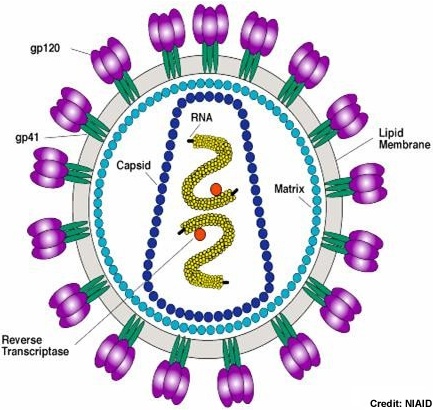
Using the viral life cycle, we talked about the various medications that are available:
  • Viral fusion inhibitors (T20)
  • Chemokine inhibitors CCR5 (miraviroc)
  • Reverse transcriptase inhibitors (NRTI -- eg tenofovir, ermtrictabine/lamivudine, abacavir) and (NNRTI -- effavirenz)
  • Protease inhibitors (lopinavir/ritonavir, atazanavir/ritonavir)
  • Integrase inhibitors (raltegravir)
General rule: 2NRTI + PI or NNRTI in treatment naive, in experienced patients who are failing goal is to develop regimen which has 3 active drugs in multiple classes.

We discussed the "AIDS" defining criteria (CD4 less than 200 or specific conditions - see list). Guidelines for the management of HIV and opportunistic infections are available here.

Monday, April 13, 2009

Day #278 - Cavitary Lung Lesion


Today we heard the case of a young man with a history of constitutional symptoms (sweats, weight loss) in association with a non-productive cough and a cavitary right upper lobe infiltrate.

I have previously blogged about the differential of cavitary lung lesions

Based on the presentation I favor an infectious etiology, most likely tuberculosis. The absence of AFB on the bronchoscopy does *not* mean this isn't tuberculosis.

This study of 230 cases of culture positive pulmonary tuberculosis showed that the BAL AFB stain was only positive in 48/95 patients without spontaneous sputum production. In this study, the sensitivity of BAL AFB stain was better, but still only 70%.

The take home message is that a negative smear does not exclude TB in a compatible case. Repeat samples are sometimes indicated and awaiting the final culture is also required. In this case I would add induced sputum daily in the AM for three days to maximize my chances of making the diagnosis.

This article reviews the various radiographic manifestations of pulmonary tuberculosis.

Treatment of Pulmonary TB (see Canadian Tuberculosis Standards):
Initial:
  • Isoniazid (INH) +/- Vitamin B6
  • Rifampin (RIF)
  • Pyrazinamide (PZA)
  • Ethambutol (ETH)
If demonstrated susceptible to INH/RIF/PZA:
  • Can stop ETH immediately
  • Continue INH/RIF/PZA until 2 months then stop PZA
  • Continue INF/RIF 4 months to complete 6 total months

Wednesday, July 22, 2009

TB vs PCP

See previous blogs on TB here and PCP here. (NB the search function is behaving a little wonky this week -- known blogger issue). Article on PCP in acute seroconversion here -- it is very rare

Friday, May 1, 2009

Day #295 - Tuberculosis and Immune Reconstitiution


We discussed a case of pulmonary and extrapulmonary tuberculosis which presented on TNF-alpha antagonist therapy.

Addendum: TB Immune Reconstitution Reviewed Recently here.

We also discussed paradoxical worsening of TB post-discontinuation of immunosuppression. The case report I was talking about is available here.

Thursday, July 31, 2008

Day #31 - Fever of Unknown Origin

Today we discussed a case of true fever of unknown origin.

Epidemiologic Criteria:
  • Fever x 3 weeks (38.3)
  • Failure to find cause despite appropriate initial outpatient/inpatient investigations
Causes:
  • Majority unknown
  • Then infection (most commonly TB or occult abscesses), inflammatory, malignancy, other

Diagnostic tests (beyond initial):
  • CT abdomen to look for abscess, lymphadenopathy, other pathology. Yield ~20%
  • Bone scan (insensitive, but specific with good + LR)
  • Liver biopsy -- regardless of hepatomegaly or LFT abnormalities has yield ~15%
  • Temporal artery biopsy in the elderly -- yield ~ 15%
  • Leg dopplers -- yield 2-6%
  • Bone marrow biopsy-- in those with cytopenias or compatable syndromes. Yield ~1%

Friday, July 18, 2008

Day #18 - Cirrhosis

Today's case was of a 52 year-old man who presented with new diagnoses of cirrhosis with ascites, portal hypertension and probable variceal bleed who also had a new diagnosis of diabetes mellitus. The discussant showed us, although in his own uniquely tangential way, the type of clinical reasoning that great physicians use in arriving at a diagnosis.

There are a number of valuable teaching points today:

1) Ascites:

Etiology
  • Elevated Hydrostatic Pressure --> CHF, constrictive pericarditis, Budd-Chiari, cirrhosis, IVC occlusion
  • Decreased oncotic pressure --> malnutrition, nephrotic syndrome, protein losing enteropathy, cirrhosis/liver failure
  • Increased fluid production in peritoneum --> infection (TB), neoplasm
Diagnosis on History/Exam
History
  • Increased abdominal girth and ankle swelling are the most sensitive (greater than 85%)
  • Past history of hepatitis or cancer are the most specific (greater than 85%)
Exam


  • Not likely if no bulging flanks, no flank dullness and no shifting dullness
  • Likely if fluid wave, shifting dullness, and peripheral edema (LR+3.8/-0.2)
2) Special Tests:

The team today also mentioned that they had performed a paracentesis in order to evaluate the ascites. This is important as paracentesis can:
  1. Provide diagnostic clues
  2. Exclude SBP
The Serum Albumin-Ascites Gradient (SAAG) is a valuable tool in deciding if the ascites is due to portal hypertension. SAAG = Serum albumin - Ascites Albumin. If <11 style="font-style: italic;">It should also be prevented in cirrhotic patients post GI bleed using TMP/SMX 1DS PO BID x 7d or CIPROFLOXACIN 500mg PO BID x 7d

The diagnosis can be made on the paracentesis:
  • PMN >250 (LR + 6 LR - 0.2)
  • WBC >1000 (LR + 9)
  • Inoculation of blood culture bottles (fill them with required volume) can improve yield of the C/S
NB: In most cases, you do not need to use FFP/Platelets pre paracentesis.

Initial treatment is with CEFTRIAXONE 2g IV q24h with coverage narrowed to the culture data. You need only treat for 5 days.

Patients should get 1.5g/kg of 25% albumin within 6h then 1g/kg on day 3.

NB: Think of missed perforation if multiple organisms grow in the culture or if the WBC count in the fluid is much greater than 1000.

Thursday, February 26, 2009

Day #231 - Cavitary Lung Lesions


Today we discussed an immunosuppressed patient with bilateral upper lobe cavitary lung lesions. The differential diagnosis for such lesions in the immunocompetent includes:


Infection:
  • TB or Atypical Mycobacterial Disease
  • Cavitary Pneumonia
    • Gram negatives (Klebsiella, E. Coli, Pseudomonas)
    • Gram positives (Staphylococcus aureus (particularly CA-MRSA), group A streptococci)
    • Anaerobes (Lung Abscess)
  • Fungal: Aspergillosis, Histoplasmosis, Coccidiomycosis, Blastomycosis, Cryptococcus
  • PCP
  • Superinfection/Colonization of an existing cavity
  • Septic pulmonary emboli with cavitation
Inflammatory:
  • Wegner's Granulomatosis
  • Rheumatoid Arthritis
  • Sarcoidosis (rare)
Vascular
  • Pulmonary Infarct
Malignancy:
  • Bronchoalveolar carcinoma
  • Bronchogenic carcinoma
  • Squamous cell cancer of lung
  • Cavitating metastases
Anotomical:
  • Cystic Bronchiectasis

Wednesday, May 20, 2009

Day #314 - Pulmonary Eosinophilia

Very complex case facilitated by an expert discussant. The key focus, other than on the case at hand was a demonstration of clinical reasoning and Bayes theorem.

We highlighted the importance of Occam's Razor -- but stressed the importance of recognizing Hickam's dictum.

I have previously blogged about pulmonary eosinophilia here (with references). In my mind, an important consideration in this patient (given travel to China) is to exclude disseminated strongyloidiasis because like infliximab related interstitial lung disease (NEJM article here) 'strongy' can appear to improve on steroids.

This is a recent review from the Lancet on TNF-alpha blockade and the interaction with tuberculosis.

NB: Review of radiographic manifestations of pulmonary TB.

Monday, April 27, 2009

Day #292 - Meningoencephalitis


Today we discussed a few issues:

  1. The developing influenza outbreak (see previous blogs on influenza). A review of the neurologic manifestations of influenza infection is available here.
  2. A case of meningoencephalitis (see TWH blog), presumably due to mumps (though I have my reservations as the parotid enlargement classically predates the encephalitis and the IgM is still pending!)
  3. TB Meningitis was discussed given the epidemiology.

Monday, May 11, 2009

Day #305 - HIV Lymphoma


Today we discussed a case of a patient with known HIV disease (CD4 300-500, not on therapy) who presented with fever, diffuse adenopathy, and splenomegaly. The presumed cause is lymphoma.


The differential diagnosis would include:

Infections:
    • HIV with high level viremia
    • Mononucleosis syndromes (EBV, CMV, acute toxoplasmosis)
    • Syphilis
    • Disseminated TB
    • In the more immunosuppressed host:
      • Mycobacterium Avium Complex
      • Bartonella Henselae
      • Disseminated fungal infection (i.e. histoplasmosis)
  • Non-infectious
    • Lymphoma
    • Multicentric Castleman's Disease
    • Sarcoidosis

I have previously blogged about HIV here, here (with cryptococcal meningitis), and here (with PCP pneumonia).

I have blogged about lymphoma including staging here.

A review of the pathology of HIV associate lymphomas is here. An article reviewing the treatment of HIV associated lymphoma is here.

Tuesday, August 5, 2008

Day #36 - Acute Confusion

Today we talked about a case of an elderly woman with an acute confusion. In approaching this problem, the history is important to exclude dementia which was sub-clinical for a long period but subsequently becomes obvious to the family.

We looked at a differential for an acute change in level of consciousness and the following general framework is helpful:

  1. Drugs/Iatrogenic
  2. Infection
    • Meningitis (bacterial, viral, TB, other -- e.g. cryptococcal, neurosyphilis)
    • Encephalitis (HSV1/2, HIV, CMV, WNV, Eastern Equine encephalitis, other)
    • Non-CNS infection with delerium
  3. Metabolic
    • Electrolytes (hyper Ca, hypo/hyper Na, hypoglycemia)
    • hypothyroidism. B12/folate not usually acute.
  4. Structural
    • Tumour
    • Bleed (SDH, SAH, ICH)
    • Other mass lesion
    • Epileptogenic focus (post ictal)
    • Stroke syndrome
    • Vasculitis
  5. End-organ Failure
    • Cirrhosis (encephalopathy)
    • CHF
    • Respiratory -- Hypoxemia/Hypercarbia
    • Renal - Uremia


We then talked a bit about aphasias because the patient had anomia with a non-fluent aphasia and inability to understand.

Aphasias
1-naming-if normal - no aphasia
2-command-

DON'T UNDERSTAND
non fluent, no repeat - global aphasia
non fl - can repeat mixed transcortical
fluent - no repeating - wernikes
- can repeat - trans cortical sensory aphasia
UNDERSTAND
non fl--no repeat - brocas
non fl - can repeat - transcortical motor
fl - no repeat - conductive
fl - repeat - no name - nominal


We then entertained the possibility of neurosyphilis or a paraneoplastic syndrome such as paraneoplastic limbic encephalitis.

Neurosyphilis can present with a variable presentation.

"Early" <>
  • asymptomatic
  • meningitis (with cranial nerve palsies)
  • meningovascular -- clinical picture of strokes, recurrent. Part of the differential for stroke in the young.
  • otic syphilis -- tinnitus, vertigo, hearing loss
  • occular syphilis -- uveitis, retinitis, vitritis
"Late" >10 years
  • General paresis (of the insane):
    P - personality changes
    A - affect changes
    R - reflexes (hyper-reflexia)
    E - Eye (argyll-robertson pupils)
    S - Sensorium - delusions, hallucinations
    I - Intellect
    S - Speech (abnormal)
  • Tabes dorsalis: sensory loss (dorsal columns), ataxia, bladder dysfunction, "lightning" pains in legs
Patients should have a positive RPR (though can be negative) and positive confirmatory test like the TPPA.

CSF should:
  1. Have a positive VDRL (specific, but not sensitive)
  2. OR Have a lymphocytic pleocytosis (WBC >20) and/or protein >500mg and positive peripheral serologies
  3. Be suspected in positive CSF FT-ABs is positive

Monday, July 21, 2008

TGH "Case of the Week" July 14-18, 2008

30F with known HIV (CD4 unknown, not on treatment) presents with acute onset fever, chills, rigors, and cough with sputum. On exam she is febrile, tachycardic, hypotensive, hypoxemic on room air requiring hi-flow oxygen to maintain saturations >90% and in moderate respiratory distress.

The chest x-ray is taken and appears below:


Questions:

1) What is the most likely microbiological diagnosis?
2) What is the differential diagnosis?
3) What tests would you order to make your diagnosis?
4) What empiric treatment would you initiate in the ED?

BONUS: What "management strategy" would you employ in the ED and what journal was the study published in?

Thank you to the people who submitted answers. The "contest" is closed for this week. The winner will be notified in person.

"Answers"

1) The most likely etiologic agent to cause a lobar pneumonia in a patient with HIV is still streptococcus pneumoniae. In fact, patients with HIV are at an increased risk of getting pneumococcal infections and should all be vaccinated with the polysaccharide vaccine.

2) The differential diagnosis includes lobar pneumonia with the other organisms of community acquired pneumonia including haemophilus influenzae, moraxella cattharalis, and Staphylococcus aureus. Of particular concern in a patient who is rapidly deteriorating is community acquired MRSA necrotizing pneumonia.

Other causes would include Legionella pneumophillia and if risk factors such as underlying structural lung disease enteric gram negative organisms.

Upper lobe pneumonia should prompt concern for tuberculosis, although in this case the acquity and sepsis-syndrome argue more strongly in favor of bacterial pneumonia.

PCP tends not to be lobar and consolidative and the classic x-ray appearance looks more like bilateral peri-hilar interstitial infiltrates.

Fungal pneumonias like cryptococcal pneumonia, blastomycosis, invasive aspergillosis are unlikely and would not generally have this radiographic appearance and acute presentation.

3) Blood cultures should be sent ASAP (ideally before antibiotics) as they may be positive in up to 25% of cases of pneumococcal pneumonia. Sputum cultures should also be sent for conventional culture, legionella culture and TB culture. I would not recommend sputum for PCP in this case.

Urinary antigen detection for pneumococcal antigen is used in some centres (not here). Urinary antigen testing for legionella can help make this diagnosis.

CD4 testing in acute illness may not be helpful as the acute illness could cause a decrease in the counts; however, it would not be unreasonable as if the CD4 count was >200 rare causes are much more unlikely.

4) In a septic patient with community acquired pneumonia you need to cover broadly for the most likely pathogens. There are many ways to do this and in some cases what you choose will depend on recent antibiotic exposure.

In this case a combination of VANCOMYCIN 1gIV q12h and LEVOFLOXACIN 750mg IV/PO q24h would be appropriate. This would cover MRSA and quinolone resistant pneumococcus, the usual pathogens including the majority of pneumococcus, unusual pathogens like legionella, and many enteric gram negatives.

BONUS: The management strategy to be employed is "Early Goal Directed Therapy in Sepsis"

Tuesday, August 18, 2009

From the Conference: Hot Papers in ID

As presented by Matthew Muller (in order of presentation):
  1. "The Animal Rule" - Raxibacumab for the Treatment of Inhalational Anthrax
  2. "The Acid Truth" - Acid suppressive medication use and the risk of hospital-acquired pneumonia
  3. "If it ain't Dutch, it ain't much" - Decontamination of the Digestive Tract and Oropharynx in the ICU
  4. "Zero Tolerance for CLI" - Chlorhexadine-Impregnated Sponges and Less Frequent Dressing Changes for Prevention of Catheter-Related Infections in Critically ill Adults: a Randomized Controlled Trial
  5. "Short Red Snappers" - Adverse Events with 4 months of Rifampin therapy or 9 months of isoniazid therapy for latent TB
  6. "Short Red Snappers (2)" - Moxifloxacin vs. Ethambutol in the initial treatment of tuberculosis: a double-blind, randomized, controlled phase II study
  7. "Maybe yes, maybe no" - Corticosteroids in the treatment of severe sepsis and septic shock in adults: a systematic review. Accompanying editorial here.
  8. "Pandemic Influenza - Much ado about nothing?" - Severe Respiratory Disease Concurrent with the circulation of H1N1

Friday, September 19, 2008

Day #80 - Diarrhea

Today we talked about a case of acute diarrhea. The discussant took us through an excellent approach to acute diarrhea. Some highlights below:

Etiology:
  • Infection (great article -- also IDSA diarrhea guidelines)
    • Viral: rotavirus, enterovirus, norovirus, etc. (CMV in immunocomprimized)
    • Bacterial: shigella, yersinia, e. coli, salmonella, campylobacter, C. difficile, TB (usually chronic), atypical mycobacterial (immunocomprimised)
    • Protozoal/Parasitic: Giardia, amebiasis, etc.
  • Inflammatory
    • UC/Crohn's
  • Ischemic
    • Small bowel -- pain out of proportion, post prandial pain
    • Large bowel -- bloody stool, colitis
  • Osmotic
    • Laxatives, chewing gum diarrhea (xylulose), sorbitol, sucrose, post-surgical "dumping"
  • Endocrine
    • Hyperthyroidism, carcinoid syndrome, VIPoma

Tuesday, May 12, 2009

Day #306 - TB Pleuritis


We discussed a great case of tuberculosis causing pleural effusion today. I have previously blogged about this before.

Previous blogs on pleural effusion here and here (including discussion of Light's Criteria and parapneumonic effusions/empyema).

Modified Light's Criteria (see review here):
  • Change LDH pleural fluid 0.45 ULN (increases sensitivity but reduces specificity)

Alternative tests:

  • LDH (0.45ULN), cholesterol (45mg/dl) and protein (29g/L) of pleural fluid, done in combination have similar performance characteristics to Light's criteria and don't require paired serum samples.